Ultrasound scan is one of the most important medical procedures today, especially when it comes to pregnancy. The process is a medical procedure that uses high-frequency sound waves to capture live images from the inside of the recipient’s body. The process, also known as sonography, has been very helpful for pregnant women as it helps detect the condition of a fetus, most importantly.
With the importance of ultrasound scan already established, this post takes a look at how much the procedure costs in Nigeria today.
How much does an Ultrasound Scan cost in Nigeria?
Ultrasound is a very common medical procedure today and can be done in many medical centres or clinics across the country. It is also one of the cheapest procedures and does not require so much.
However, there are certain factors to put in mind that might significantly affect how much the process goes for. One of these factors is the part of the body you want to scan. Another is type of clinic or hospital you are looking to undertake the procedure. Government-owned hospitals are generally cheaper than private hospitals and clinics. Hence, you can expect a significant difference in price between the two.
As it stands, the ultrasound Scan can be done for anything between N5,000 and N20,000 depending on the clinic or hospital you are using and the part of the body you’re scanning. It should also be noted that encompassing drugs that would be given after the ultrasound scan could further vary the total amount you will spend during the process.
What You Should Know About Ultrasound Scan
Ultrasonography is a method of body imaging that uses sound waves to expedite making a medical diagnosis. A professional ultrasound technician is able to see inside the body using the process to answer questions that could be asked by the health practitioner caring for the patient. Generally, a radiologist will supervise the ultrasound scan test and reports to the medical doctor.
Some other types of doctors may also use ultrasound scan as a diagnostic tool. For instance, obstetricians use ultrasound scan to assess the foetus during pregnancy. Surgeons and emergency doctors use ultrasound at the bedside to evaluate abdominal pain or other similar concerns.
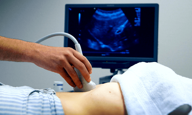
During the process, a transducer is used to project and collect the sound waves and their echoes. A gel is spread onto the patient’s skin so that the sound waves are not inaccurate as they move through the skin. Using their knowledge of human anatomy and the scanning machine, the specialist can assess specific structures and answer the question asked by the patient’s physician.
The process may take a significant amount of time and may require the transducer to be shifted and pointed in different directions. Also, the specialist may need to vary the quantity of pressure used to push the probe into the skin.
Generally, an ultrasound allows the doctor to see problems with organs, vessels, and tissues without necessarily requiring to make an incision. Unlike most other imaging methods, ultrasound does not involve radiation. Because of this, it rates among the most preferred method for viewing a developing foetus during pregnancy.
How to Prepare for an Ultrasound Scan
Usually, the essential steps you will take to get yourself ready for an ultrasound largely depend on the area or part of your body that is being examined. Your medical doctor may tell you to avoid eating anything for about 8 to 12 hours before your ultrasound scan, especially if your abdomen is being observed. There is a possibility that undigested food can block the sound waves, which makes it difficult for the specialist to get a clear picture.
For an inspection of the gallbladder, liver, pancreas, or spleen, the doctor might instruct you to eat a fat-free meal the night before the scan and then avoid eating any other food until the procedure. Nonetheless, you can carry on to drink water and take any medicines as instructed. For other analyses, you may be required to drink a lot of water and to hold your urine so your bladder could be full to further enable better visualized.
It is also important for you to inform your doctor about any prescription drugs, over-the-counter medicines, or herbal supplements that you take before the period or are currently taking. It is highly important to follow your doctor’s directives and ask as many questions you may want to ask before the procedure. There are no serious risks when it comes to ultrasound. Unlike X-rays or CT scans and most other scans, ultrasounds does not involve radiation.
Uses of Ultrasound Scans
Ultrasound is generally used for diagnosis, for treatment, and for supervision during techniques such as biopsies. The process can also be used to scan internal organs such as the liver, kidneys, the pancreas, the thyroid gland, the testes and the ovaries, and many other parts of the body.
An ultrasound scan can disclose whether a lump is a tumor. This could be tumorous, or a fluid-filled cyst. It can also help diagnose issues with soft tissues, muscles, blood vessels, tendons, and joints. It is used to examine a frozen shoulder, tennis elbow, carpal tunnel syndrome, and others.
Doppler ultrasound, a type of ultrasound, can help assess the flow of blood in a vessel or blood pressure. It can define the speed of the blood flow and any obstacles. An echocardiogram, also known as ECG, is a popular example of a Doppler ultrasound. It can be used to generate images of the circulatory system and to measure blood flow and cardiac tissue movement at intervals.
Related Stuff
- Best Hospitals for Kidney Transplant in Nigeria (2024)
- Cost of HSG Test in Nigeria (April 2024)
- Cost of IVF in Nigeria (2024): All You Need to Know
- Cost of Pap Smear in Nigeria (April 2024)
- The Cost of IUI in Nigeria (2024)
- The Cost of Kidney Transplant in Nigeria (2024)
- Fohow Products Price List (April 2024)
- Cost of DNA Test in Nigeria (April 2024)
- Cost of Fibroid Surgery in Nigeria (April 2024)
- Cost of Treating Hepatitis B in Nigeria (2024)
- Cost of Tuberculosis Test in Lagos Nigeria (April 2024)
- The Cost of Hysteroscopy in Nigeria (2024)
- Sphygmomanometer Prices in Nigeria (April 2024)
- Synlab Price List (April 2024)
- Cost of Endoscopy in Nigeria (April 2024)
- Glucometer Prices in Nigeria (April 2024)
- Cost of Kuding Tea in Nigeria (2024)
- Cost of Radiotherapy in Nigeria (April 2024)
- Cost of Dialysis in Nigeria (April 2024)
- Afriglobal Medicare Price List (April 2024)
- Cost of Laparoscopy in Nigeria (April 2024)
- Cost of Medical Exam for Canada Immigration in Nigeria
- Cost of MRI in Nigeria (April 2024)
- Nordica Fertility Centre Price List (April 2024)
- Me Cure Nigeria Price List (April 2024)
- Cost of Ultrasound Scan in Nigeria (April 2024)
- Cost of Chemotherapy in Nigeria (2024)
- Cost of PCR Test in Nigeria (April 2024)
- Omron Blood Pressure Machine Prices in Nigeria (2024)
- Cost of Dental Braces in Nigeria (April 2024)
")
")
")
")
: All You Need to Know")
")